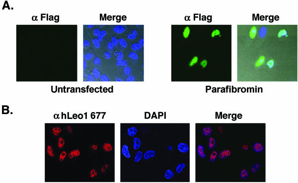
FIG. 4.
Parafibromin and hLeo1 are nuclear proteins. (A) Immunofluorescence assay with FITC-conjugated anti-Flag antibody on untransfected HeLa cells (Untransfected) or HeLa cells transfected with Flag-tagged WT parafibromin (Parafibromin). The left side of each pair of images shows the FITC signal, while the right side, marked “Merge,” is the integration of the FITC and DAPI signals overlaid upon the Nomarski-differential interference contrast images of the cells. (B) Immunofluorescence with anti-Leo1 antibody Ab677 (Cy3 red) was used to visualize endogenous Leo1 (left image), and DAPI staining (middle image) was used to mark nuclear DNA in HeLa cells. The right image, labeled “Merge,” shows the colocalization of the two signals.

