Figure 1.
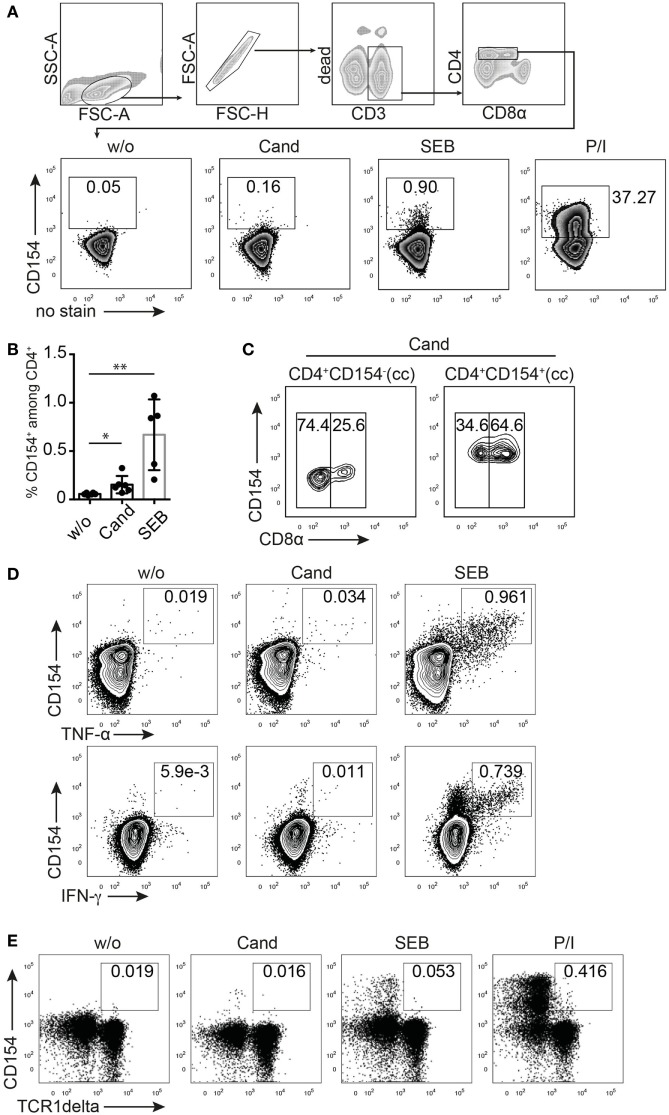
CD154 identifies porcine, antigen-reactive CD4+ T cells. T cell receptor stimulation (TCR)-independent and TCR-dependent activation induced CD154 expression in pig CD4+ T cells detected by flow cytometry and intracellular staining. (A) Gating strategy and representative zebra plots showing the CD154 signal from ex vivo PBMC gated on CD4+ T cells that were either unstimulated (w/o) or stimulated with Candida albicans lysate (Cand, 40 µg/ml), staphylococcal Enterotoxin B (SEB, 1 µg/ml), or PMA/ionomycin (P/I) for 6 h. Panel (B) summarizes w/o, Cand, and SEB stimulatory conditions for PBMC from n = 5 to 6 animals (w/o vs. Cand, p = 0.0271, Student’s t-test and w/o vs. SEB, p = 0.0043, Mann–Whitney test). (C) Analysis of CD8α coexpression of Candida-reactive CD154+CD4+ T cells or non-responding CD154−CD4+ T cells after Cand stimulation of PBMC. Concatenated contour plots (from n = 3 animals) are illustrated and numbers in gates identify CD8α negative (left rectangle gate) or positive (right rectangle gate) CD4+ T cells. (D) Flow cytometry of CD154/Cytokine coexpression analysis in either Cand (40 µg/ml) or SEB (1 µg/ml) stimulated PBMC for TNF-α, IFN-γ, and interleukin-17A. (E) Analysis of CD154 expression in gamma delta T cells (identified by TCR1δ+ expression and pregated on live CD3/duplet exclusion/FSC-SSC properties) upon antigen-specific (Cand, 40 µg/ml), superantigen-specific (SEB, 1 µg/ml), and TCR unspecific (PMA/ionomycin) stimulation.

