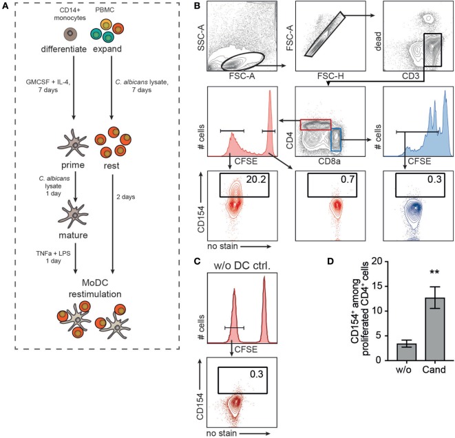
Figure 2.
Pig antigen-responding T cells can be expanded and visualized after monocyte-derived dendritic cell (MoDC) restimulation. (A) Experimental setup. Purified blood CD14+ monocytes were differentiated into MoDC for 7 days in the presence of GM-CSF and interleukin-4. MoDCs were primed with 40 µg/ml Candida albicans lysate and maturation was triggered by TNF-α and LPS treatment for 1 day. In parallel, PBMCs were CFSE-labeled and expanded in the presence of 20 µg/ml Candida antigen for 7 days followed by a 2-day resting phase. Expanded lymphocytes were restimulated with autologous, primed MoDC (MoDC:T cell ratio, 1:5) and CD154 expression was assessed according to the gating strategy in (B). CD154 expression of CD4+CFSElow (proliferated cells, lower left plot) was compared to unproliferated CD4+CFSEhigh (lower middle, red plot) and proliferated CD4−CD8α+CFSElow T cells (lower right, blue plot). (C) As control, CD154+ frequency of Cand expanded PBMC without DC restimulation (w/o DC ctrl.) is shown exemplarily [gated on CD4+CFSElow as depicted in panel (B)]. (D) CD154+ T cell frequencies after restimulating expanded T cells with unprimed (w/o) or Candida antigen-primed (Cand) MoDC (n = 6, Mann–Whitney test, **p = 0.0043).

