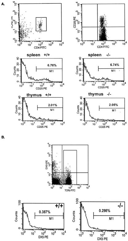
FIG. 4.
(A) The percentages of thymic and splenic CD4+CD25+ T cells were not affected by deletion of Prss16. Splenic and thymic cells of Prss16+/+ and Prss16−/− mice were stained with anti-TCRβ, anti-CD4, and anti-CD25. The percentages of TCRβ+CD4+ cells expressing CD25 are indicated. (B) NK T cells identified as TCRβ+DX5+ from spleens of Prss16+/+ and Prss16−/− mice were analyzed by using three-color FACS analysis. The numbers indicate the percentages of DX5+ T cells.

