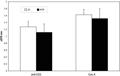
FIG. 5.
Activation of splenocytes by anti-CD3 MAb and concanavalin A from Prss16−/− mice is not different from that from control littermates. Splenocytes were treated for 48 h with either anti-CD3 MAb or concanavalin A, and proliferation was assessed by an MTT assay. Bars represent the means ± standard deviations of the differences in absorbance at 570 nm between anti-CD3-, concanavalin A-, and PBS-treated cells. Each group of 10 mice was assayed in triplicate.

