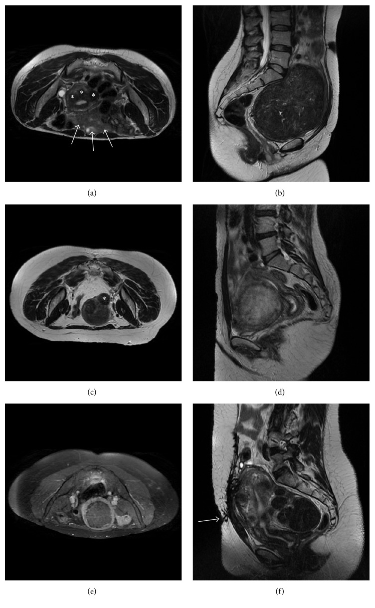
Figure 1.
Illustrative pelvic MRI scans of excluded patients: (a) axial T2-w showing three small fibroids (asterisks) and many bowel loops (white arrows) that are interposed between the skin surface and the hypothetic target; (b) sagittal T2-w of a large fibroid which almost occupies the whole pelvis and is dangerously close to sacrum bone and nerves (this patient performed the screening MRI in supine feet first position since she reported some discomfort in maintaining the prone position); (c) axial T2-w showing a small and pedunculated subserosal fibroid (asterisk); (d) sagittal T2-w of a “bright” untreatable cellular fibroid; (e) axial T1-w with fat saturation acquired after intravenous injection of paramagnetic contrast medium showing a nonenhancing fibroid; (f) sagittal T2-w of a patients with a bulky scar in abdominal skin (white arrow).

