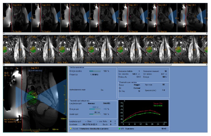
Figure 2.
Screenshot from the ExAblate workstation showing the planned target of patient in case 3 after the first sonication performed. In bottom left, the HI-FU beam representation is shown in light blue, the red line indicates the skin-gel pad interface, and critical structures are secured by the use of specific low-energy density region (LEDR) and no-pass regions markers (bowel in pink, pubic bone in light blue). The target volume (region of treatment, ROT) has been split into multiple subvolumes (green and yellow voxels) each of which will be ablated by a specific sonication. Patients' movements during treatment are monitored by reviewing fiducials (red crosshairs) placed by the treating physician on distinct anatomic structures which can be monitored during real-time MR imaging. Real-time thermal mapping after the first sonication is shown in the bottom right graph (maximum temperature achieved in the focal spot is 71°C).

