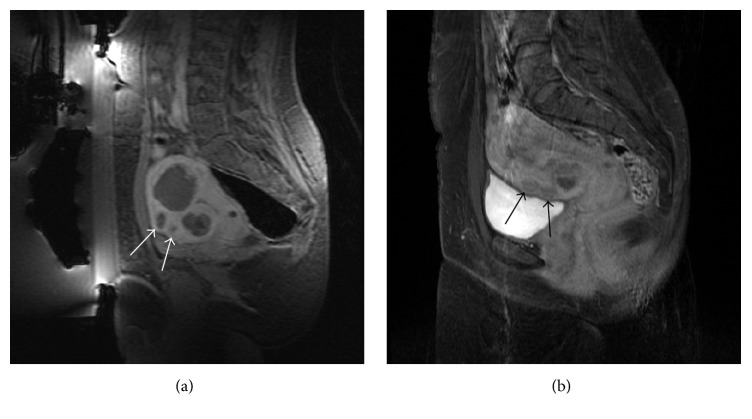
Figure 3.
Case 1: (a) sagittal T1-w with fat saturation acquired after intravenous injection of paramagnetic contrast medium showing the two small fibroids (white arrows) located in the near field that were not directly treated but that became nonperfused too after the treatment; two bigger fibroids treated are visible too; (b) a follow-up MRI (T1-w with fat saturation acquired after intravenous injection of paramagnetic contrast medium) showing a normal vascularization of the two small fibroids.

