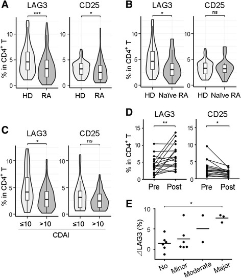
Fig. 3.

Frequencies of LAG3+ Tregs and CD25+ Tregs in patients with RA and in healthy volunteers, as well as the effect of abatacept treatment. a and b PBMCs were taken from healthy donors (HD; n = 101) and patients with RA (n = 85, including 16 nontreated patients [naive RA]), and Treg subsets were assessed. c Patients with RA were divided into two groups according to Clinical Disease Activity Index (CDAI). Treg subsets were assessed (CDAI ≤10, n = 30; CDAI >10, n = 55). d Treg subsets were analyzed immediately before and 6 months after abatacept treatment (n = 18). e Changes of percentages of LAG3+ Tregs in CD4+ T cells (ΔLAG3) were evaluated in accordance with treatment response to abatacept (n = 18). No response was defined as less than 50% improvement from baseline CDAI. Minor response was defined as at least a 50% improvement from baseline CDAI. Moderate response was defined as at least a 70% improvement from baseline CDAI. Major response was defined as at least an 85% improvement from baseline CDAI. *, P < 0.05; **, P < 0.01; ***, P < 0.001. Statistics: (a–c) unpaired two-tailed Student’s t tests, (d) Wilcoxon signed-rank test, (e) Kruskal-Wallis test and Dunn’s multiple-comparisons test. CD Cluster of differentiation, LAG3 Lymphocyte activation gene 3, ns Not significant, PBMC Peripheral blood mononuclear cell, RA Rheumatoid arthritis, Treg Regulatory T cell
