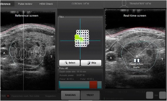
Fig. 1.

A picture of the touch-screen interface of the HIFU device. The central panel represents the birdview reconstruction of the nodule made out of multiple white cycles. The empty circles represent the unablated subunits while the filled circles represent the ablated subunits. The hyperechoic marks, on the right, are a sign of tissue necrosis from the ablationᅟ
