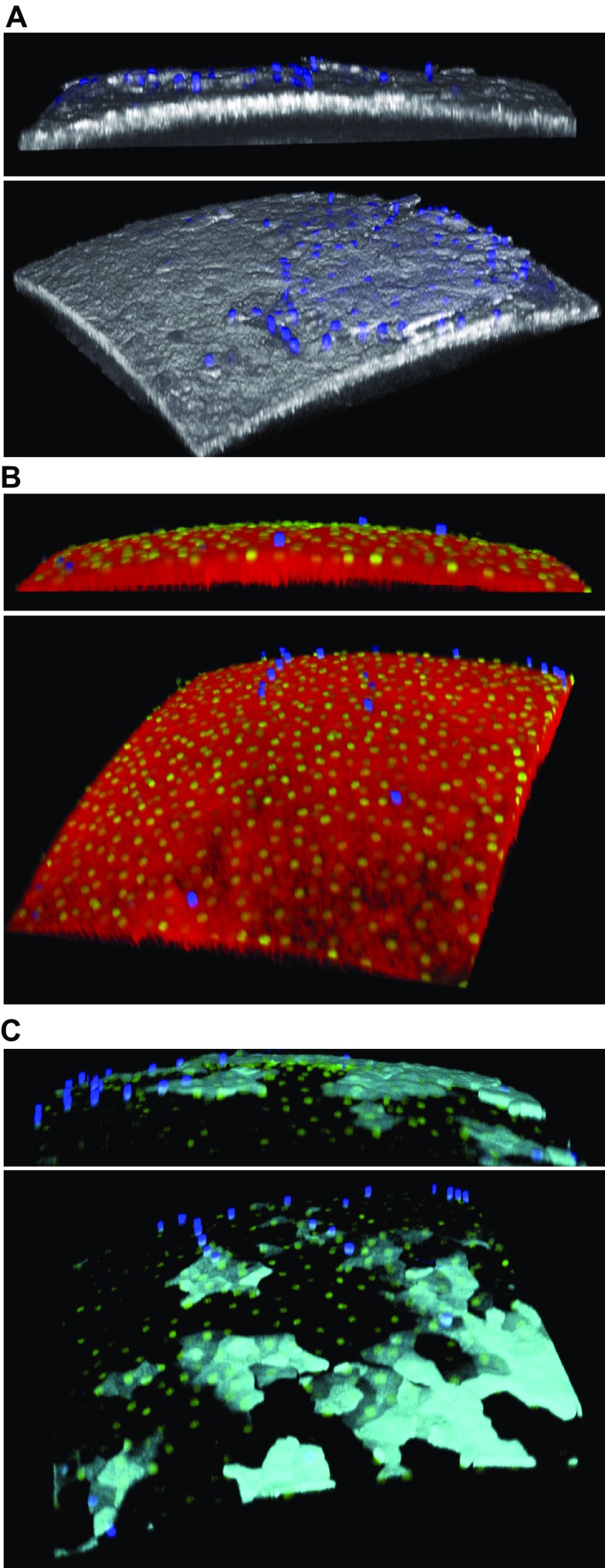
Figure 2.
Murine corneal epithelial cell health after overnight (24 h) Ac4GalNAz incubation ex vivo followed by DIBAC-biotin and streptavidin–Alexa Fluor 488 labeling. A) Reflectance image (white) after additional DAPI labeling shows several exfoliating dead cells (blue). B) Live/dead stain of epithelial surface (reflectance in red). DAPI was added to incubation medium to show dead cells (blue), and SYTO Green 16 dye was added to show nuclei of live cells (green). C) GalNAz labeling (cyan) merged with DAPI and SYTO Green 16 images revealed live and dead cells colocalizing with sugar label. In total, 3 eyes from 3 WT mice were analyzed in 2 separate assays using live/dead dyes after GalNAz labeling; representative images are shown.

