
Figure 3.
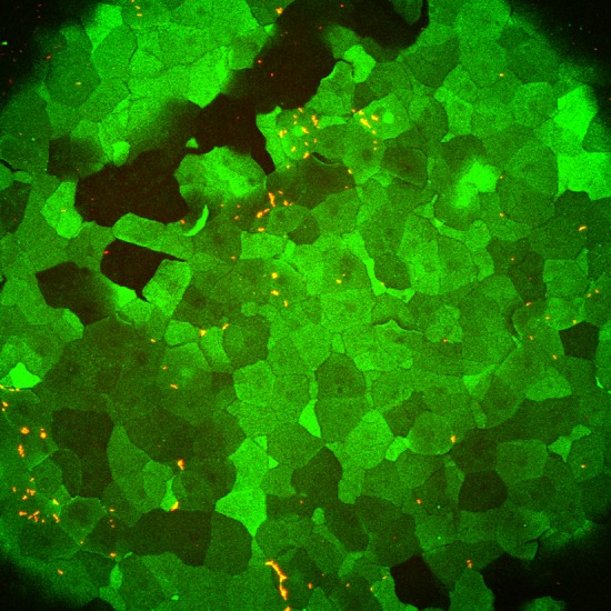
P. aeruginosa colocalization with GalNAz-labeled cells on murine cornea ex vivo. Approximately 5 × 10⁸ CFU of P. aeruginosa (dTomato, red) was added for 1 h to cornea that was previously incubated with Ac4GalNAz overnight before washing the eye and incubating with DIBAC-biotin and streptavidin–Alexa Fluor 488. Representative image is shown. Three WT mouse eyes were quantified to determine degree of association between bacteria and GalNAz-labeled cells (see Results). Surfaces were generated in Imaris software to determine bacterial colocalization in 3 dimensions.