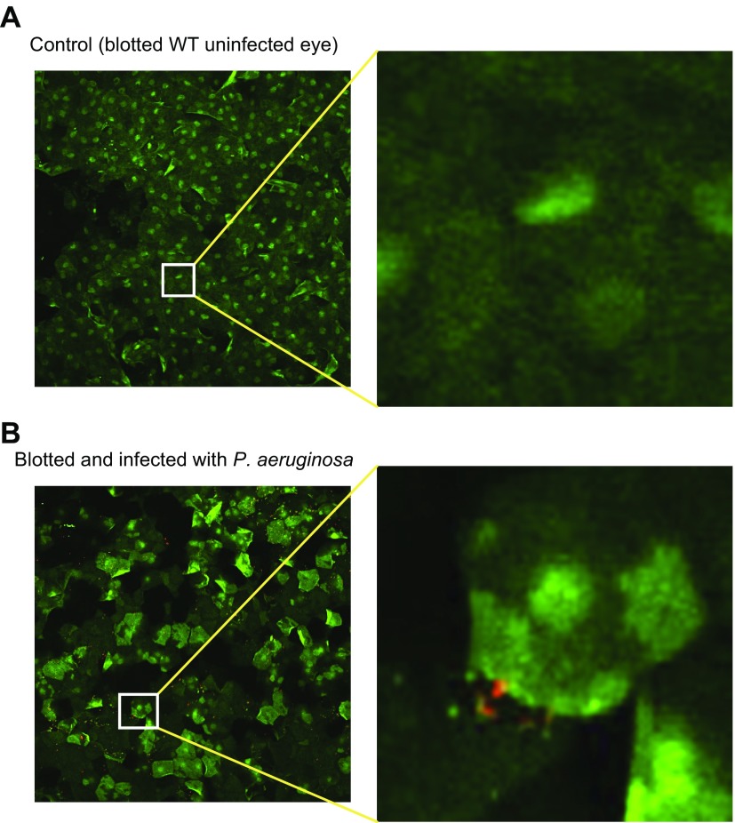
Figure 6.
P. aeruginosa enhances GalNAz labeling after blotting. WT mouse eyes were incubated in Ac4GalNAz overnight, blotted, and then after 10 min incubated in control MEM (A) or ∼5 × 10⁸ CFU P. aeruginosa (B) for 1 h before addition of DIBAC-biotin and streptavidin–Alexa Fluor 488. GalNAz-labeled mucins (green) are shown as maximum intensity projection; they appear upregulated and redistributed in blotted corneas incubated with bacteria compared to medium-only control (original magnification, ×20 for both corneas). Surfaces were generated in Imaris software to determine bacterial colocalization in 3 dimensions. Images are representative of 3 mouse corneas (A) and 5 mouse corneas (B) analyzed in 2 separate assays.

