Figure 1.
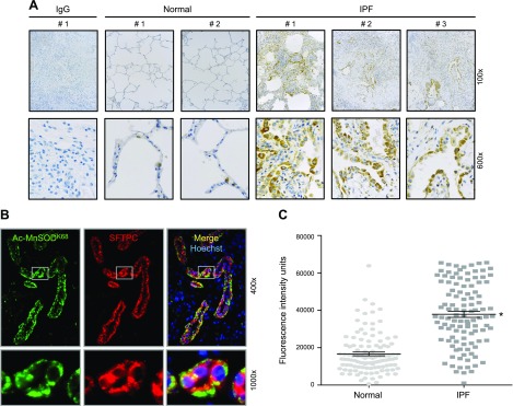
Tissue from patients with IPF has increased acetylation of MnSODK68, a known SIRT3 deacetylase target, which is localized to AT2 cells. A) Explanted lungs from patients with end-stage IPF were evaluated via IHC by using Abs that were targeted to a negative control IgG (fibrotic lung) and Ac-MnSODK68 from normal controls and fibrotic lungs. Shown is a representative panel of 2 normal and 3 fibrotic lungs (for the remainder, please see Supplemental Fig. 1). B) Dual immunofluorescence (IF) staining was used to colocalize expression of acetylated MnSODK68 (green fluorescence) and SFTPC (red fluorescence, used as a marker for AT2 cells). C) Semiquantitative analysis, measured by per-cell fluorescence intensity units as described in Materials and Methods, was used to compare AcMnSODK68 expression in AT2 cells of fibrotic (n = 110) and normal control (n = 121) lungs. Bars shown indicate means ± sem. *P < 0.05 vs. normal AT2 cells.

