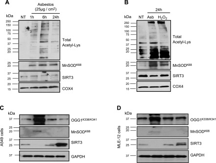
Figure 3.
Total AEC mitochondrial protein, OGG1, and MnSOD show increased acetylation after oxidative stress or SIRT3 silencing. A, B) MLE-12 cells (A) were exposed to asbestos (25 µg/cm2) for variable periods, and primary isolated rat AT2 cells (B) were exposed to asbestos (25 µg/cm2) or H2O2 (200 µM) for 24 h, with mitochondrial protein isolated. Global mitochondrial protein lysine acetylation, acetylated MnSODK68, SIRT3, and GAPDH expression were assessed by Western blotting. Shown are representative blots from a total of 3 experiments. C, D) A549 (C) and MLE-12 (D) cells were transfected with siRNA that was targeted to SIRT3 (siRNA) or enforced expression of a SIRT3 WT plasmid (SIRT3-WT) for 48 h, then total cellular protein was extracted and expression of acetylated OGG1K338/K341, acetylated MnSODK68, SIRT3, and GAPDH was assessed by Western blotting. Shown is a representative blot from a total of 3 experiments. COX4, cytochrome oxidase IV; NT, no treatment.

