Figure 3.

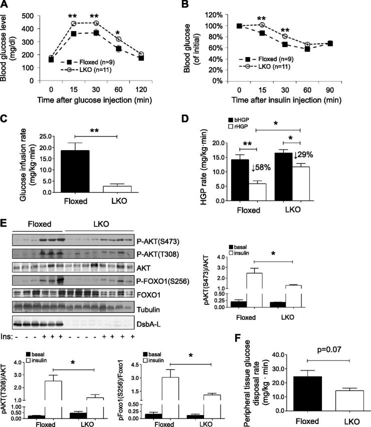
Liver-specific knockout of DsbA-L exacerbated diet-induced insulin resistance. A, B) Glucose tolerance test (A) and insulin tolerance test (B) were performed on DsbA-LLKO (LKO) and floxed control mice fed with HFD for 8 wk. C) Glucose infusion rate of DsbA-LLKO and floxed control mice fed with HFD for 8 wk was measured by hyperinsulinemic–euglycemic clamp studies (n = 4 per group). D) HGP under basal (bHGP) and during hyperinsulinemic–euglycemic clamp (rHGP). E) HFD-fed DsbA-LLKO and floxed control mice were unfed overnight before intraperitoneal injection of insulin (1 U/kg). Mice were humanely killed 15 min after insulin injection, and tissues were dissected on ice and frozen in liquid nitrogen until homogenization. Insulin-stimulated phosphorylation of PKB and Foxo1 in liver of DsbA-LLKO and floxed control mice was determined by Western blot analysis. F) Peripheral tissue glucose disposal during hyperinsulinemic–euglycemic clamp. All values represent means ± sem. *P < 0.05, **P < 0.01 (1-way ANOVA).
