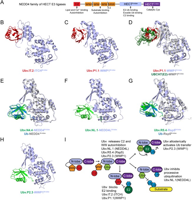
Figure 3.

Ubv inhibitors, activators, and modulators of HECT E3 ligases. (A) Domain composition of the NEDD4 subfamily of HECT E3 ligases. (B) and (C) Structures of Ubv inhibitor complexes: Ubv.IT2‐ITCH (PDB: 5C7M) (B) and Ubv.P1.1‐WWP1 (PDB: 5HPS) (C). Only the N‐lobe of HECT domains is shown. Ubv and HECT N‐lobe are colored red and light blue, respectively. (D) Superposition of Ubv.P1.1‐WWP1 and UBCH7‐WWP1 (PDB: 5HPT) complexes showing that Ubv inhibitors target the E2 binding surface. Structural alignment was produced by aligning WWP1 HECT N‐lobe domains. The Ubv.P1.1‐WWP1 complex subunits are colored as in (B) and the UBCH7‐WWP1 complex subunits are colored as follows: WWP1 N‐lobe, grey; UBCH7, dark green. (E), (F), (G), and (H) Structures of Ubv activator/modulator complexes: Ubv.N4.4‐NEDD4 (PDB: 5CJ7) (E), Ubv.NL1.1‐NEDD4L (PDB: 5HPK) (F), Ubv.R5.4‐Rsp5 (PDB: 5HPL) (G), and Ubv.P2.3‐WWP1 (PDB: 5HPT) (H). Only the N‐lobe of HECT domains is shown. Ubv and HECT N‐lobe are colored green and light blue, respectively. Ubv.N4.4‐NEDD4 (E) and Ubv.R5.4‐Rsp5 (F) complexes are shown superimposed with Ub.wt‐NEDD4 (PDB: 4BBN) or Ub.wt‐Rsp5 (PDB: 3OLM), respectively. Structural alignment was produced by aligning N‐lobe of HECT domains. Subunits of Ub‐NEDD4 and Ub‐Rsp5 complexes are colored as follows: Ub.wt, blue; HECT N‐lobe, grey. (I) Schematic of HECT3 ligase activation and catalysis depicting observed roles of different Ubvs
