Fig. 5.
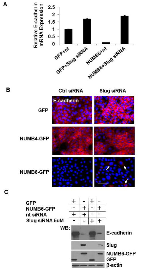
Slug knockdown partially restores E-cadherin expression in NUMB6-GFP DB-7 cells. Western blot analysis, real-time PCR and immunostaining assays were carried out to evaluate the expression of E-cadherin in DB-7 cells overexpressing control vector with GFP or NUMB6-GFP. (A) Control vector GFP and Numb-6 GFP DB-7 cells were transfected with control siRNA or Slug siRNA for 48 h and then E-cadherin levels were evaluated by qPCR. NUMB6-induced loss of E-cadherin mRNA expression was recovered when cells were treated with Slug siRNA. (B) Control GFP, NUMB4-GFP or NUMB6-GFP DB-7 cells were transfected with control siRNA or Slug siRNA, respectively. Immunostaining was performed in 48 hours later to assess the expression levels of E-cadherin; red is Alexa Fluor-546 for E-cadherin; nuclei were stained with DAPI. Bars=15um (C) Control GFP, or NUMB6-GFP DB-7 cells were transfected with control siRNA or Slug siRNA, and E-cadherin expression was evaluated by Western Blot.

