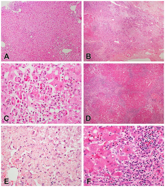
Figure 5.
Necrosis patterns in DILI. A, C, E. Zonal necrosis due to mithramycin (A, C) and acetaminophen (E). In both of these examples there is some combination of coagulative necrosis and apoptosis in zone 3 without much inflammation beyond an infiltrate of macrophages. In the case of acetaminophen, the coagulative necrosis mainly affects the hepatocytes as the sinusoidal lining cells remain intact (E). B, D, F. Sub-massive necrosis due to isoniazid injury. The necrosis in this case is irregular, with large areas of complete multiacinar necrosis (B) next to areas that show some necrosis along with regenerative nodules (D). Inflammation remains in residual portal areas and along the edges of the regenerative nodules (F)

