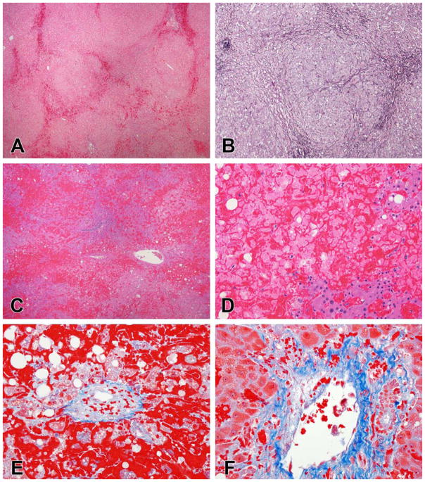
Figure 9.
Examples of vascular injury. A., B. Nodular regenerative hyperplasia due to oxaliplatin injury. There is congestion of dilated sinuses between the regenerative nodules (A). The reticulin stain shows the irregular liver cells plates more clearly, with widened, 2-cell thick plates of enlarged hepatocytes within the nodules and compressed plates of atrophic hepatocytes between the nodules (B). C, D, E, F. VOD/SOS following hematopoietic stem cell transplant. There is extensive hemorrhage and necrosis (C), with only a few groups of residual hepatocytes (D), mainly in zone 1. Trichrome stains show that the central veins are narrowed by loose, pale staining connective tissue (E). Some of the portal veins also showed partial occlusion with loose connective tissue (F).

