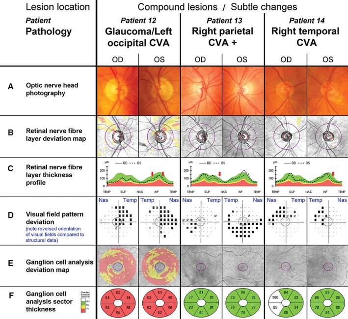
Figure 5.

Common, non‐classical clinical findings with retrograde degeneration. The central grey circle in each of the visual field pattern deviations (D) corresponds to the area assessed by the ganglion cell analysis (E, F). D. Please note that conventionally visual field test results are aligned to display the left eye on the left side and the right eye on the right hand side of the observer. As indicated by the blue labels, the opposite order was chosen for this figure to align visual field tests with all other results of the respective eye, resulting in a reversal of the orientation for this panel only. Additional patient details are provided in Table 2 and in the text. Patient 12. A–C. Patient 12 had bilateral thin superior neuroretinal rims and beta parapapillary atrophy paralleled in the bilateral arcuate superior nerve fibre defects (B, C). D. The patient's visual field revealed right superior homonymous quadrantanopia. There was superior depression in both eyes and additional inferior nasal depression in the left eye. E, F. Generalised ganglion cell layer thinning was present. Patient 13. A–F. Patient 13 presented with left inferior quadrantanopia, extending temporally in the left eye (D) but no other noticeable abnormalities in the optic nerve head or ganglion cell analysis. Patient 14. A–F. Patient 14 displayed a visual field defect consistent with slightly incongruous left superior quadrantanopia (D), which was not accompanied by any other noticeable changes in the optic nerve head or ganglion cell analysis (A–C, E, F). It should be noted that the right temporal ganglion cell layer appeared thicker than normal (F).
