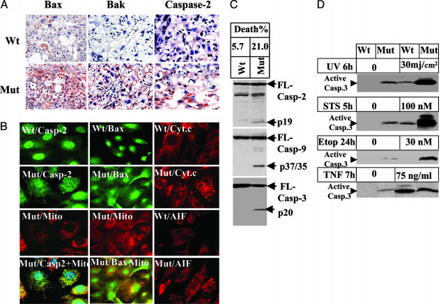
Fig. 3.
Ablation of Bruce activates the mitochondrial pathway of apoptosis. (A) Immunohistochemical staining of placentas with antibodies against Bax, Bak, and caspase-2. (B) Subcellular localization of proapoptotic factors in Birc6GT MEFs. Mitochondria were stained with Mito Tracker red. Caspase-2, Bax, and cytochrome c were stained by their primary antibody followed by Alexa Fluor 488-conjugated secondary antibody (green) for caspase-2 and Bax, and by Texas red for cytochrome c and AIF (red). The yellow fluorescence in the overlay of caspase-2 or Bax with Mito Tracker indicates mitochondrial colocalization of the two molecules. Smear staining of cytochrome c in the mutant MEFs indicates the release of cytochrome c. Nuclear staining of AIF indicates translocation of AIF to the nucleus in mutant MEFs. (C) Apoptosis in MEFs. Fifty micrograms of protein extract was assayed by Western blot for cleavage of caspase-2 (Top), caspase-9 (Middle), and caspase-3 (Bottom). The spontaneous death rate of Birc6GT mutant MEFs was 21%, as determined by propidium iodine staining and scored for subdiploid DNA contents by flow cytometry analysis. (D) Sensitivity of MEFs to apoptotic stimuli. MEFs were subjected to UV, staurosporin, etoposide, or TNF plus 0.5 μg/ml cycloheximide. Cell death was scored by Western blot for the activation of caspase-3.

