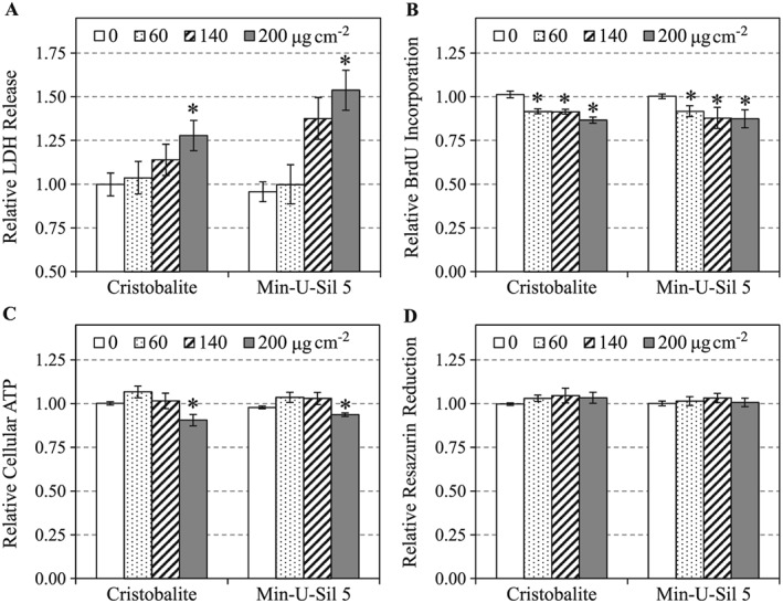
Figure 2.

Cytotoxicities of cristobalite and Min‐U‐Sil 5 in A549 cells after 24 h of exposure were assessed by LDH release (A), BrdU incorporation (B), cellular ATP (C) and resazurin reduction (D) assays. Data are expressed as mean fold effect ± standard error, relative to control (0 μg cm–2), n = 4. Two‐way ANOVA was used to determine significant effects of the particles, where Holm–Sidak was the post‐hoc method used for all pairwise comparison procedures. *Significant change (P < 0.05) compared to control (0 μg cm–2). BrdU, 5‐bromo‐2′‐deoxyuridine; LDH, lactate dehydrogenase.
