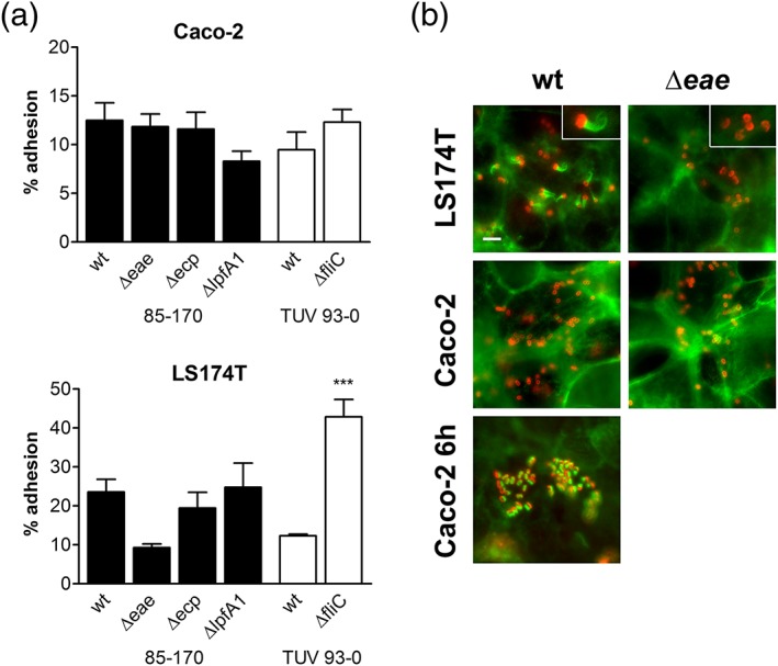
Figure 2.

Involvement of EHEC adhesins in binding to Caco‐2 and LS174T cells. (a) Adherence of wild‐type (wt) and adhesin‐deficient EHEC strains after 3 hr of infection. Adhesion was determined by colony‐forming unit counting and is expressed as percentage of cell‐bound bacteria relative to the inoculum. ***p < 0.001 versus wt. (b) Fluorescent actin staining to identify A/E lesion formation. Caco‐2 and LS174T cells were infected with EHEC 85‐170 wt or Δeae for 3 or 6 hr (Caco‐2 for 6 hr) and stained for actin (green) and E. coli (red). Inserts in top right corner show enlarged image areas containing EHEC bacteria with and without actin pedestals (LS174T wt and Δeae, respectively). Bar = 5 μm
