
Figure 4.
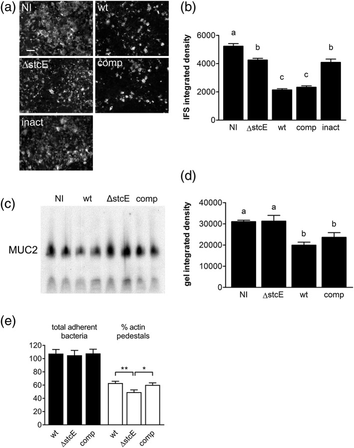
EHEC StcE reduces mucin levels and promotes A/E lesion formation on LS174T cells. Cells were infected with wild‐type TUV 93‐0 (wt), an isogenic stcE mutant (ΔstcE), ΔstcE complemented with wild‐type (comp), or catalytically inactive StcE (inact), or left non‐infected (NI) for (a–d) 6 hr or (e) 3 hr. (a) MUC2 expression was determined by immunofluorescence staining, bar = 5 μm or (c) agarose gel electrophoresis and Western blotting and quantified by integrated density measurement (b and d, respectively). Means with different letters are significantly different (p < 0.001 for b, p < 0.01 for d). n = 2 in duplicate for c and d. (e) A/E lesion formation was assessed by immunofluorescence staining and expressed as percentage of bacteria associated with actin pedestals relative to the total number of adherent bacteria, **p < 0.01, *p < 0.05