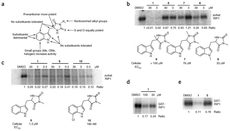
Figure 2.

SAR analysis of RIP1 inhibition by necrostatins. (a) Summary of cellular SAR of 1 based on ref. 14. (b,c) In vitro activity of select inactive (b) and active (c) 1 analogs. Endogenous RIP1 kinase assays were performed as in Figure 1e in the presence of indicated amounts of 1 analogs. Structures of the derivatives are shown. EC50 values for inhibition of cellular necrosis in TNFα-treated FADD-deficient Jurkat cells by necrostatins were determined as described in the Methods and were previously reported14. (d,e) 1 inhibits kinase activity of recombinant RIP1 expressed in Sf9 cells. Recombinant RIP1, expressed in Sf9 cells, was subjected to in vitro kinase assay in the presence of indicated amounts (d) or 100 μM (e) of 1 (d,e) or 5 (e). Assays were performed at least two or three times, and similar results were obtained each time. The representative images are shown.
