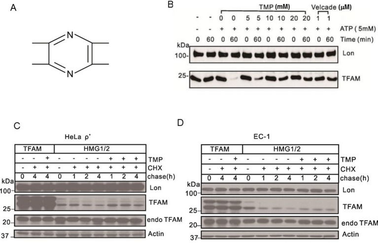
Figure 1. TMP blocks Lon-mediated TFAM degradation.
(A) Chemical structure of TMP. (B) Western blot showing levels of Lon and TFAM. Purified Lon (300 nM) was reacted with purified TFAM (150 nM) in the presence of ATP (5 mM) for 1 h. Velcade was used as the positive control. (C) Western blot showing levels of Lon, actin and TFAM. HeLa ρ+ cells were transfected with plasmids for expressing TFAMHMG1/2 or TFAMwt transiently. After transfection, cells were treated with TMP (10 μM) while chased by CHX (100 μg/ml) for the next 4 h. (D) Western blot showing levels of Lon, Actin and TFAM. EC-1 cells were transfected with plasmids for expressing TFAMHMG1/2 or TFAM transiently. After transfection, cells were treated with TMP (10 μM), while chased by CHX (100 μg/ml) for the next 4 h (endoTFAM, the endogenous TFAM).

