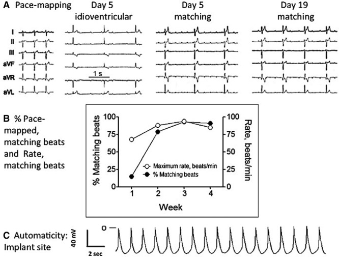
Figure 5.

A, Representative ECGs recorded during pace mapping at time of implant and 5 and 19 d after embryoid bodies (EBs) implantation. B, Time course of the percent and maximum rate of matching beats recorded from the same dog during 4 wk of follow-up. C, Automatic rhythm recorded from a ventricular slab removed from the site of EB injection on the day of the terminal study.
