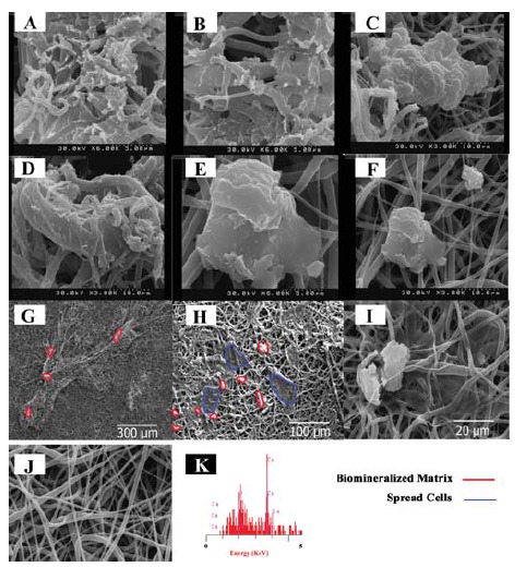
Figure 2 .

SEM images from the cellular scaffolds for calcium deposition after 7 days culture, A, B: The middle layers of the construct in dynamic culture; C, D: The layers were directly exposed to the dynamic culture medium (upper and lower layers of the construct); E, F: Static culture of the monolayer cellular scaffolds; G: Upper layer of the construct in large scale; H: Upper layer of the construct in large scale; I: Static culture of the monolayer cellular scaffolds in large scale; J: The electrospun scaffold before seeding; K: Energy-dispersive X-ray (EDX) from sample A
