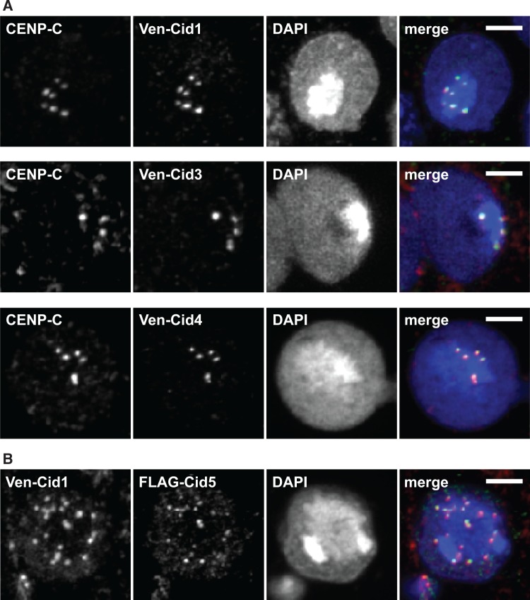
Fig. 4.
Proteins encoded by Cid paralogs localize to centromeres in cell culture. (A) Venus-tagged D. auraria Cid1, Cid3, and Cid4 were transiently transfected in a D. auraria cell line (top, middle, and bottom panels, respectively). Cells were fixed and co-stained with a D. melanogaster CENP-C antibody (red in merged image) and anti-GFP (green in merged image). These data show co-localization of all three montium subgroup Cid proteins with CENP-C. (B) We co-transfected Venus-tagged Cid1 and FLAG-tagged Cid5 from D. virilis into a D. virilis cell line. Venus-Cid1 (red in merged image) and FLAG-Cid5 (green in merged image) both formed co-localized foci in the nucleus. All scale bars indicate a distance of two microns.

