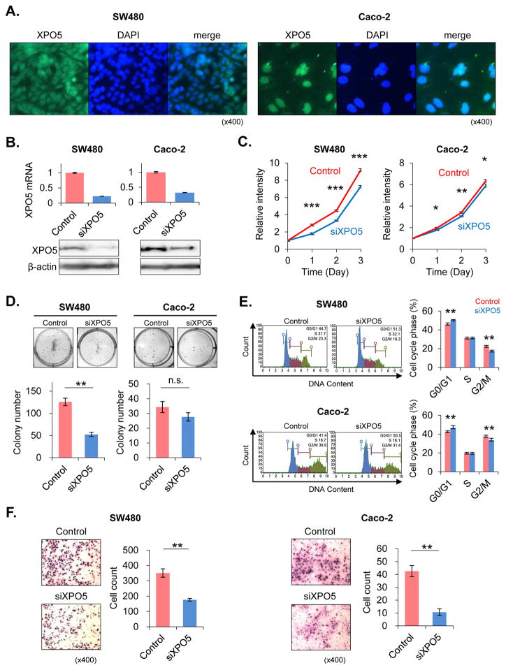
Fig. 3. Knockdown of XPO5 in colorectal cancer cells.
(A) XPO5 was detected in nuclear compartment in SW480 and Caco-2 cells in immunofluorescence staining. (B) XPO5 was knocked down in SW480 and Caco-2 cell lines using siRNA. The XPO5 knockdown level was analyzed using real-time PCR. XPO5 expression levels in XPO5 siRNA (siXPO5)-transfected cells were <30% of the levels measured in control siRNA (siControl)-transfected cells. The knockdown effect was also analyzed using western blotting. In each cell line, XPO5 protein expression was significantly suppressed by siXPO5 knockdown. (C) In the SW480 cell line, siXPO5-transfected cells exhibited significantly suppressed proliferation compared to siControl-transfected cells (P<0.0001). Similarly, XPO5 suppression resulted in reduced proliferation in Caco-2 cells (P=0.0489). (D) Colony-formation assays were performed to examine the effect of siXPO5 knockdown on the colony-forming abilities of single cells plated in vitro. siXPO5-transfected SW480 cells formed a significantly fewer colonies than siControl-transfected cells (P=0.0039). The same tendency occurred in Caco-2 cells, although this result was not significant. (E) Cell cycle analyses revealed a significant increase in the G0/G1 phase fraction after siXPO5 knockdown in both SW480 and Caco-2 cells (P=0.0090 SW480; P=0.0090 Caco-2). (F) To determine whether XPO5 knockdown inhibited cell invasion, in vitro invasion chamber assays were performed. In both SW480 and Caco-2 CRC cells, siXPO5 transfection significantly reduced invasiveness compared to siControl-transfected cells (P=0.0088 SW480; P=0.0088 Caco-2). *P<0.05, **P<0.01, ***P<0.001

