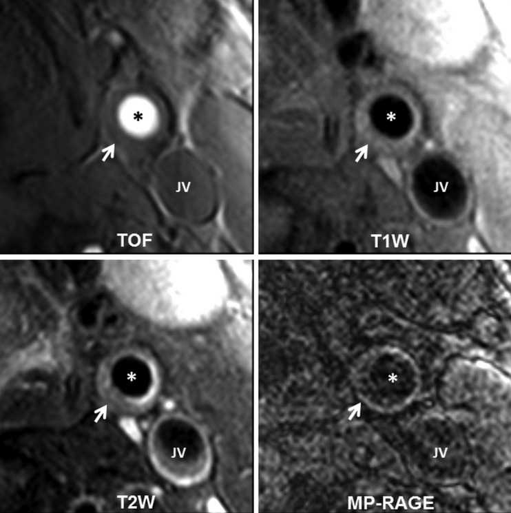
Figure 3.
Example of 2D multicontrast carotid vessel wall imaging protocol including TOF, T1W, T2W and MP-RAGE sequences. The lumen (*) and outer wall boundaries are well delineated on vessel wall images. A lipid-rich atherosclerotic lesion can be seen in the left common carotid artery (arrow). JV represents jugular vein. 2D, two-dimensional; MP-RAGE, Magnetisation Prepared Gradient Recalled Echo; TOF, time-of-flight; T1-W and T2-W, T1 and T2-weighted image.

