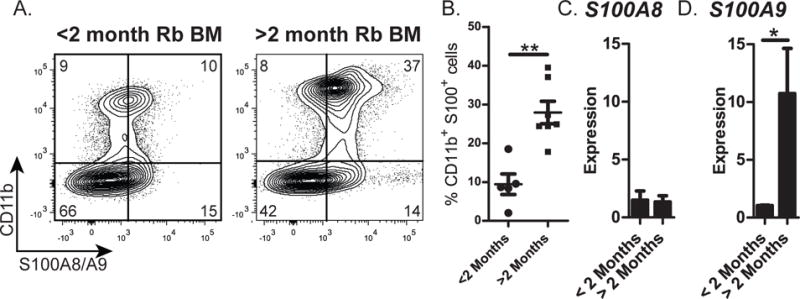
Figure 6. Quantitation of S100A8 and S100A9 in BM of rabbits before and after the arrest of B lymphopoiesis.

Rabbit BM nucleated cells were obtained from the adipocyte-free pellet and analyzed for the expression of S100A8 and S100A9. (A) Representative flow cytometry profiles of BM cells stained for the expression of CD11b (surface) and S100A8/A9 (intracellular). (B) Percentage of CD11b+S100A8/A9+ cells in BM of <2-month-old and >2-month-old rabbits. Data were from n=12. Error bars represent the average ±SD. (C and D) qRT-PCR of BM cells from rabbits >2- and < 2-months-of-age for expression of (C) S100A8 and (D) S100A9. Expression was normalized to HGPRT housekeeping gene. Data are the average from three independent experiments. Error bars represent the average ±SD. (B-D) Statistical significance was determined by Student’s t test.
