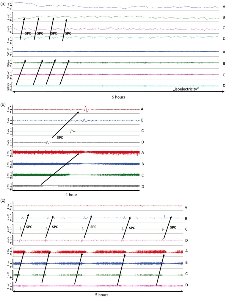
Figure 2.
Types of SDs: (a) ECoG indicates clusters of “ISDs” (n = 18 per 5 h) in a patient with significant PHE progression 24 h after hematoma evacuation; (b) Isolated spreading depolarization starting over channel D and spreading to channel A (c) ECoG during 5 h indicates clusters of stereotyped spreading depressions starting at channel D and spreading to channel A. ECoG: electrocorticography; ISD: isoelectric depolarization; SD: spreading depolarization; PHE: perihematomal edema.

