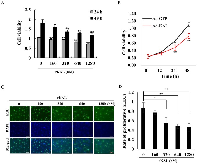
Figure 1.
Kallistatin inhibits proliferation of lymphatic endothelial cells. (A) Cells in different groups were treated with various concentrations of rKAL for 24 and 48 h. After treatment with CCK8, changes in the optical density (OD) value at 450 nm in a micro-plate reader were recorded. (B) Cells were transfected with Ad-GFP/Ad-KAL for various periods of time, then the OD values at 450 nm were recorded. (C) Cells in different groups were treated with various concentrations of rKAL for 24 h, during which cells were incubated with EdU, then tested with immunofluorescence; scale bar represents 100 µm. (D) Histogram representing the rate of proliferative hLECs. *,#P<0.05, **,##P≤0.01, the results are presented as the mean ± standard deviation.

