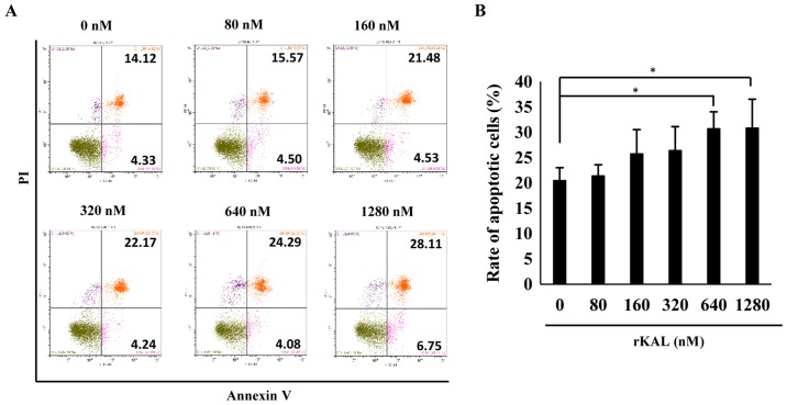
Figure 2.
Kallistatin promotes apoptosis of lymphatic endothelial cells. (A) Flow cytometric analysis of cell apoptosis in hLECs treated with various concentrations of rKAL for 48 h. (B) Histogram representing the apoptotic rate of hLECs. *P<0.05, **P<0.01, the results are presented as the mean ± standard deviation.

