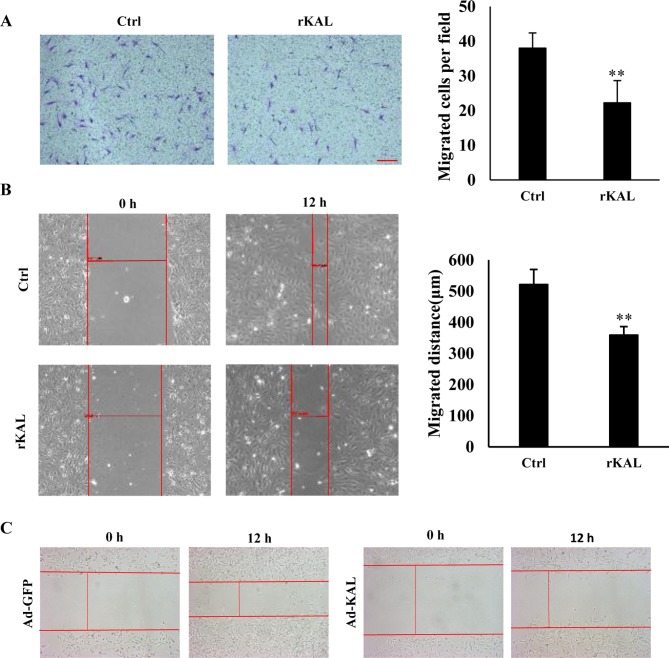
Figure 3.
Kallistatin inhibits hLEC cell migration. (A) hLECs were treated with 640 nM rKAL or PBS in a Boyden chamber assay for 12 h, then stained with crystal violet; histogram represents the rate of migration; scale bar represents 100 µm. (B) At 100% confluence, a 10-µl pipette was used to create a wound in the layer of hLECs, then cells were treated with rKAL for 12 h and microscopically imaged; migration distances were automatically measured by the software; the histogram represents the migration distance. (C) hLECs were transfected with Ad-GFP/Ad-KAL for 48 h, then distances were measured as described above. *P<0.05, **P<0.01. Results are presented as the mean ± standard deviation. hLECs, human lymphatic endothelial cells.

