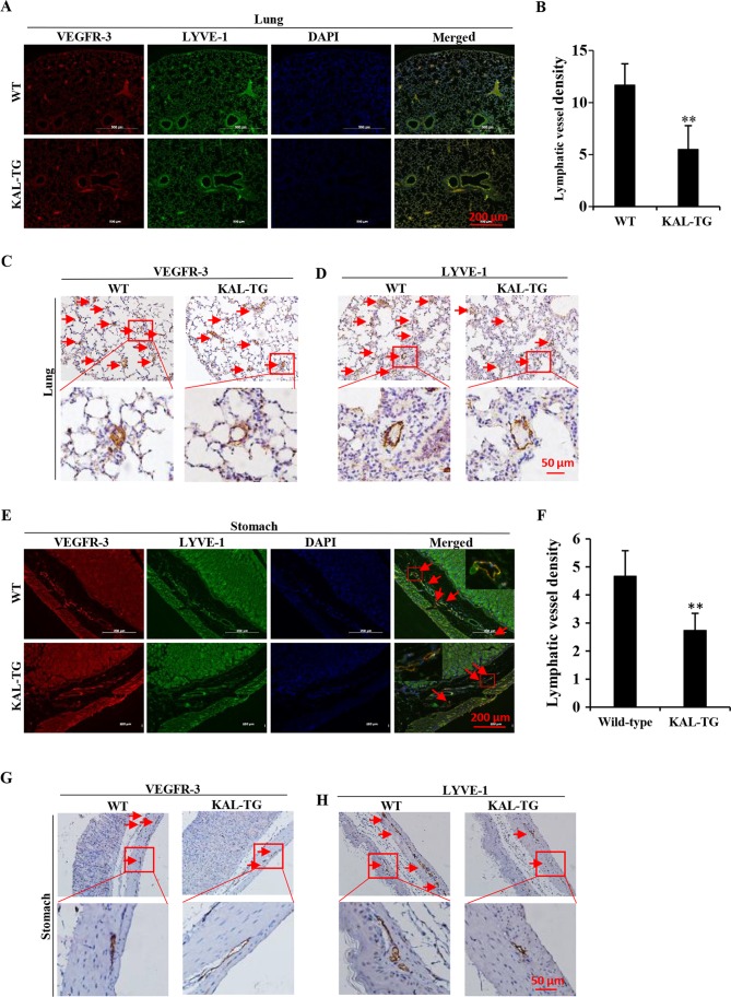
Figure 5.
The lymphatic vessel density (LVD) in different tissues of kallistatin transgenic mice (KAL-TG). (A, C and D) LVD in the lung tissue of wild-type and KAL-TG mice. The lymphatics were stained with VEGFR-3 and LYVE-1. (B) The number of lymphatics per field in lung sections (3–6 fields were counted in each group). (E, G and H) LVD in the stomach of wild-type mice and KAL-TG mice. The lymphatics were stained with VEGFR-3 and LYVE-1. (F) The number of lymphatics per field in stomach sections. n=5, wild-type and KAL-TG mice. Scale bar, 100 µm. *P<0.05, **P<0.01. Results are presented as the mean ± standard deviation.

