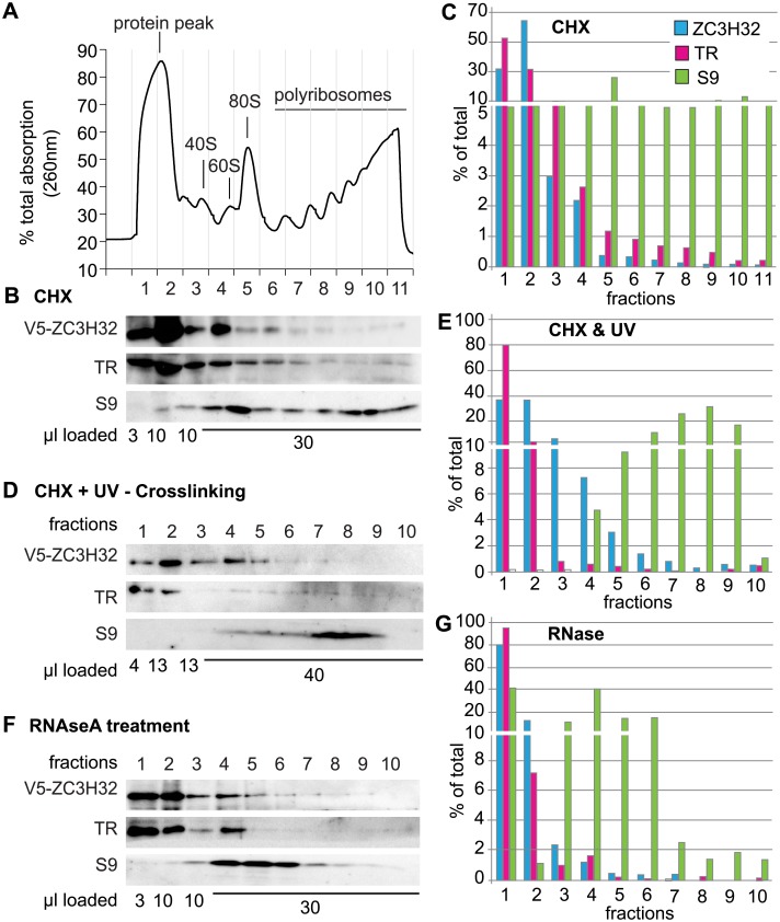
Fig 2. No evidence for association of ZC3H32 with polysomes.
(A) A typical optical density profile after sucrose density gradient centrifugation of extracts from trypanosomes expressing V5-tagged ZC3H32, and treated briefly with cycloheximide. Fraction numbers are below; the heaviest fraction is fraction 11. (B) Typical Western blot showing the distribution of V5-ZC3H32 for (A), trypanothione reductase (TR) and ribosomal protein S9. (C) Quantitation of (B), with results adjusted for loading. Note the interruption of the scale. (D) As in (B), but the trypanosomes were UV-irradiated prior to lysis in order to bind proteins to RNA. 10 fractions were obtained. (E) Quantitation of (D)—as in C. Results are also similar to (C). (F) As (B) but 100 μg/ml RNase was included in the polysome buffer. (G) Quantitation of (F).

