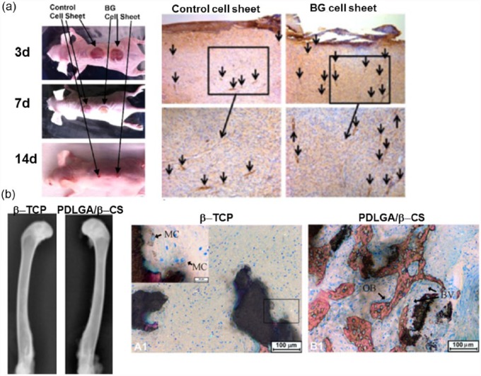
Figure 4.
In vivo angiogenesis stimulated by silica-based biomaterials in (a) wound healing model: wound closure at different time points and CD31 staining for the new blood vessels (arrows) at day 14 and in (b) bone defect model: β-TCP and PLDLA/β-CS implantation after 4 weeks, radiographs and Van Gieson’s picrofuchsin staining performed (BV: blood vessel; MC: multinucleate cell, and OB: osteoblast cell). Reprint permission was obtained from Wang et al.15 and Yu et al.56

