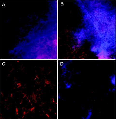
FIG. 6.
Micrographs of pellicle and biofilm aggregates stained with the red fluorescent DNA stain propidium iodide and the blue β-glucan stain calcofluor. (A and B) Ech 3937 and WPP67 pellicles, respectively. In both cases, fibrous β-glucan-stained fibers are visible surrounding the cell aggregates. (C) Solid surface-associated biofilms from WPP96 did not stain with calcofluor. (D) β-Glucan-stained fibers were visible surrounding cell aggregates in some smears from WPP101 cultures.

