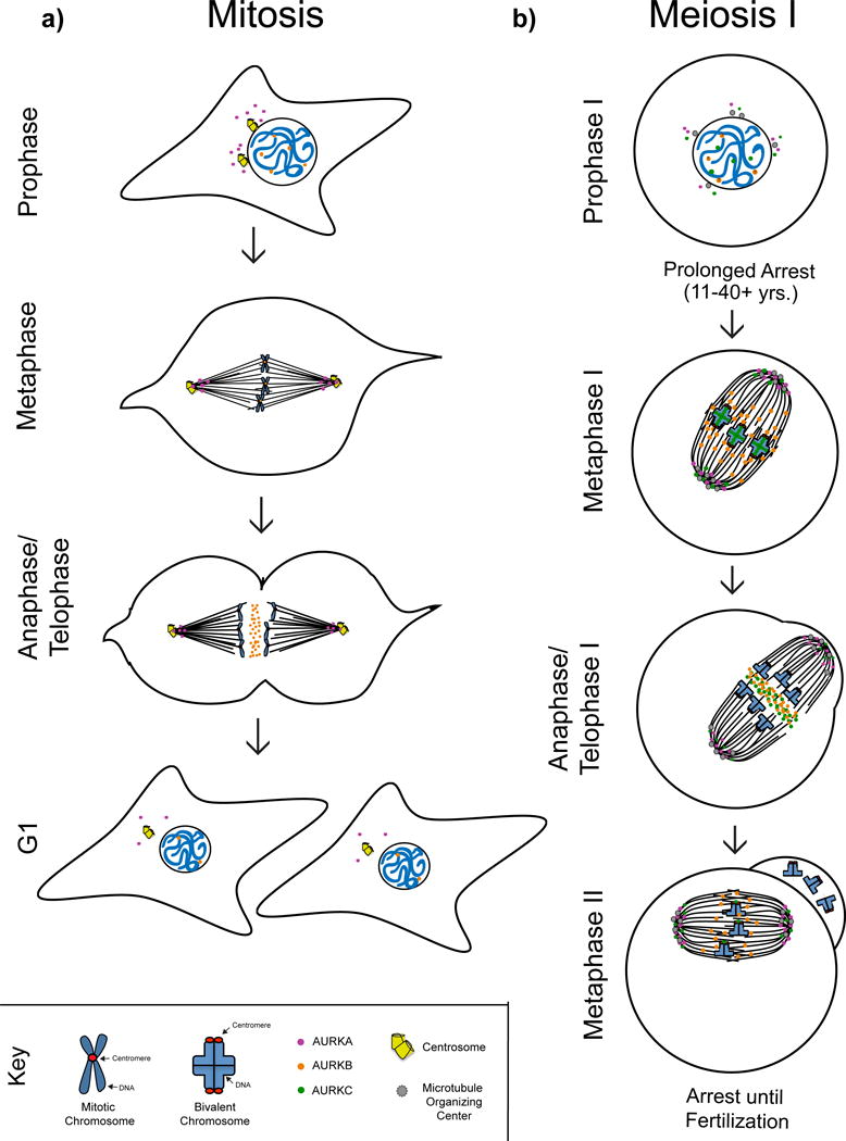
Figure 2. Aurora kinase localization in mitosis and meiosis.

Schematic representation of AURKA, AURKB, and AURKC localization in mitosis and meiosis in mammalian cells. a) In mitotic prophase AURKA (purple circles) is concentrated around duplicated centrosomes while AURKB (orange circles) is nuclear. In metaphase AURKA is found at spindle poles while AURKB is located on centromeres. In anaphase AURKA remains at spindle poles whereas AURKB concentrates at the spindle midzone. Daughter cells enter G1 stage where the expression of both kinases is substantially reduced. b) In prophase of meiosis I AURKA clusters around microtubule organizing centers in the cytoplasm while the location of AURKB remains unknown. AURKC (green circles) can be found both in the nucleus and cytoplasm. In metaphase I AURKA localizes to spindle poles while AURKB is on spindle microtubules and potentially kinetochores. AURKC is found both at spindle poles and at the interchromatid axis of MI bivalents. In anaphase I AURKA and AURKC localize to spindle poles while AURKB and a subset of AURKC concentrate at the spindle midzone. Mammalian oocytes arrest at metaphase of meiosis II until fertilization with AURKA and AURKC at spindle poles, AURKB on the spindle microtubules and AURKC concentrated at the centromere.
