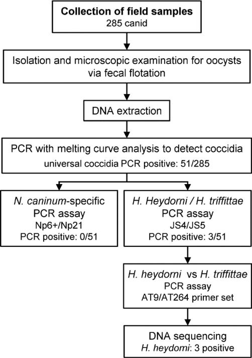
Fig. 1.
Study design flow chart. Collection of 285 canid fecal samples from the field, followed by fecal flotation for microscopic examination and isolation of parasite ova. DNA isolated from flotation material with subsequent PCR testing, first with a broad universal coccidia PCR assay whereby the 51 positive samples were further analyzed using Neospora caninum and Hammondia PCR assays with DNA sequencing of the three Hammondia heydorni positives.

