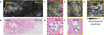
Fig. 3. Imaging of a breast tumor from the first patient.

(A) UV-PAM image of the fixed, unprocessed breast tumor. (B) H&E-stained histologic image of the same area shown in (A) acquired after processing, sectioning, and staining the excised breast tissue. The blue dashed lines in (A) and (B) outline the interface between the normal and tumor regions. (C and D) Zoomed-in UV-PAM and H&E-stained images of the red dashed regions in (A) and (B), respectively. (E and F) Zoomed-in UV-PAM and H&E images of the yellow dashed regions in (A) and (B), respectively. IDC, invasive ductal carcinoma; DCIS, ductal carcinoma in situ. (G) Zoomed-in UV-PAM image of the orange dashed region in (A). CN, cell nuclei.
