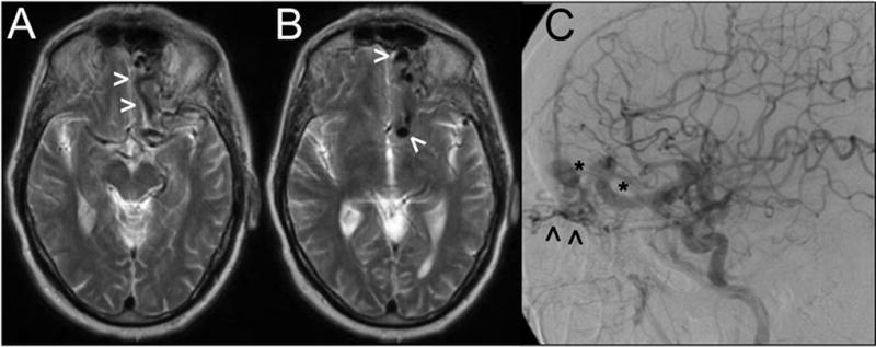
Figure 3.

Axial sections from a T2-weighted MRI study (A, B) showing large vascular flow voids (arrowheads) near the left anterior fossa floor. Digital subtraction angiography (lateral view following a left CCA contrast injection) demonstrates a Borden-Shucart Type III dAVF fed by ethmoidal branches from the ophthalmic artery (arrowheads) and draining into enlarged, arterialized cortical frontal veins with an associated venous ectasia and varices (asterisks).
