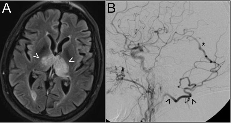
Figure 4.

This patient presented with rapidly-progressive dementia. Axial MR imaging with FLAIR sequences (A) demonstrates bi-thalamic edema (arrowheads) as a result of venous hypertension. Digital subtraction angiography (B; lateral view following a left CCA contrast injection) shows a high-grade dAVF fed by the occipital artery (arrowhead) with early arteriovenous shunting into the deep venous system (e.g., vein of Galen, asterisk). Endovascular embolization resulted in complete fistula obliteration and resolution of clinical and radiographic abnormalities.
