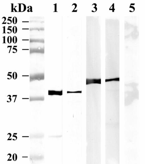
FIG. 2.
Western blot of pull-down assays using recombinant GST-LcrH-2 and His-LcrE. Molecular mass markers are shown on the left. Purified GST-LcrH-2 is shown in lane 1 and was visualized using anti-GST MAb. Lane 2 contains the eluate from a His-LcrE Ni-NTA affinity column to which GST-LcrH-2 was applied and the column was washed and eluted with imidazole and reacted with anti-GST MAb. Purified His-LcrE is shown in lane 3, visualized using anti-His MAb. Lane 4 contains the eluate from a GST-LcrH2 affinity column to which His-LcrE was applied and the column was washed and eluted with glutathione and reacted with anti-His MAb. Lane 5 shows the negative control for lane 2, where GST alone was passed through a His-LcrE affinity column and the column was washed and eluted with imidazole and incubated with anti-GST MAb.

