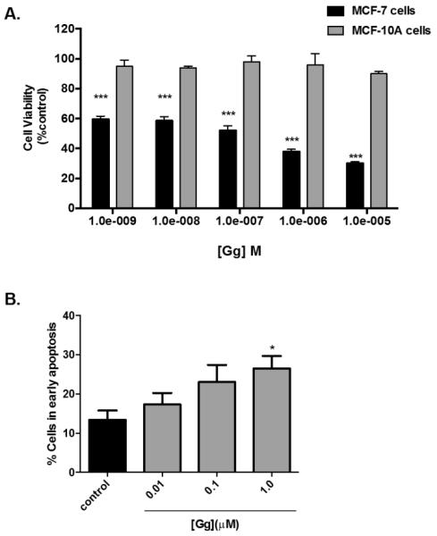
Figure 3.

Gg displays no appreciable cytotoxicity in MCF-10A breast epithelial cells and apoptosis in MCF-7 breast cancer cells.(A) MCF-10A were exposed to Gg (1 nM-10 μM) 4 OH-Tam (1 nM-10 μM) or vehicle for 72 h before Alamar Blue™ assay analysis was employed, as outlined in Materials and Methods. Statistical significance as indicated by *** P < 0.001 versus vehicle control.(B) MCF-7 cells were exposed to media containing Gg (0.01-1.0 μM) or 0.025% DMSO for 24 h before being analysed for apoptosis using the AnnexinV-7AAD assay, as described in Materials and Methods. Data represent the mean percentage ± SEM of three independent experiments performed in triplicate. Statistical significance as indicated by * P < 0.05 versus vehicle control.
