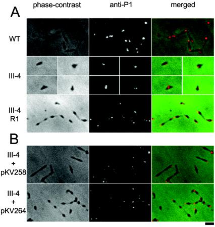
FIG. 4.
Phase-contrast, anti-P1 immunofluorescence, and merged micrographs of M. pneumoniae. (A) Wild-type (WT), mutant (III-4), and revertant (III-4-R1). Due to the poor growth of mutant III-4 and its sparse distribution on the slide, individual cells are shown in separate panels. (B) Mutant III-4 transformed with pKV258 or pKV264. Bar, 2 μm.

