. 2017 May 8;9(2):160–168. doi: 10.4055/cios.2017.9.2.160

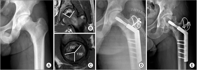
Fig. 1. A 35-year-old man had osteonecrosis in the left femoral head. (A) Preoperative anteroposterior hip radiograph. (B and C) The combined necrotic angle was 240°. (B) The angle of the necrotic area in the mid-coronal image was 90°. (C) The angle of the necrotic area in the mid-sagittal image was 150°. The angle between the central vertical line of the femoral head and the posterior margin of the necrotic portion was 135°. (D) Immediate postoperative anteroposterior hip radiograph after transtrochanteric rotational osteotomy. (E) Follow-up radiograph taken 6 years after the operation showing osteophyte formation around the femoral head.

Fig. 1